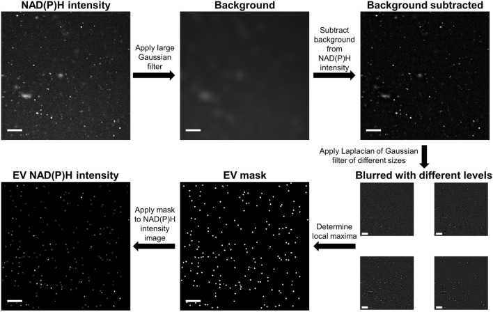
Figure 2.
Schematic of extracellular vesicle (EV) segmentation using a blob detection algorithm. First, a large Gaussian filter was applied and then subtracted from the original intensity image to adjust for nonuniformity in the background. Next, Laplacian of Gaussian filters of different sizes were applied to the image to highlight local maxima of varying sizes. A 3D local maxima filter was applied to a stack of images filtered at different levels to create a mask for EVs of different sizes and a threshold was applied to only segment maxima that exceed the background noise. Finally, this binary mask was multiplied by the original intensity image to display the intensity of each segmented EV. Scale bars represent 20 μm.

