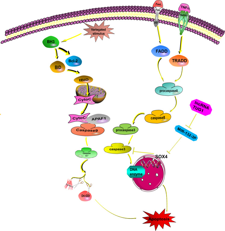
Fig. 1. Activation of the apoptotic pathway.
Apoptosis can be divided into the extrinsic pathway and intrinsic pathway. In the extrinsic pathway, the death receptor TNFR, Fas/CD95, and TRAIL combine with the relevant factors from extracellular, such as TNF, FasL. Then, the death receptor, procaspase-8, caspase-8, and caspase-3 are sequentially activated and regulate DNA fragmentation factor (DFF)/caspase-activated DNase (CAD) and cause apoptosis26,27,36. In the intrinsic pathway, cytotoxic stimuli such as carcinogenic stress, chemotherapeutic agents, and developmental cues activate BH3-only family members and inhibit Bcl-2 proteins, thus activating the pro-apoptotic effectors BAX and BAK, which finally destroy the outer mitochondrial membrane. This releases cytochrome C and ATF endonuclease. Cytochrome C engages APAF1 and forms the apoptosome, which activates caspase-9. Subsequently, caspase-9 activates caspase-3, caspase-6, and caspase-7, which contribute to the cleavage of other proteins and eventually promote apoptosis. Various lncRNAs are involved in this process. For instance, TUG1 can inhibit miR-132-3p expression, thereby increasing SOX4 expression. This decreases caspase-3 activity, eventually inhibiting osteosarcoma (OS) cell apoptosis. This indicates that knockdown of TUG1 could regulate the miR-132-3p–SOX4 axis in OS cells and promote apoptosis.

