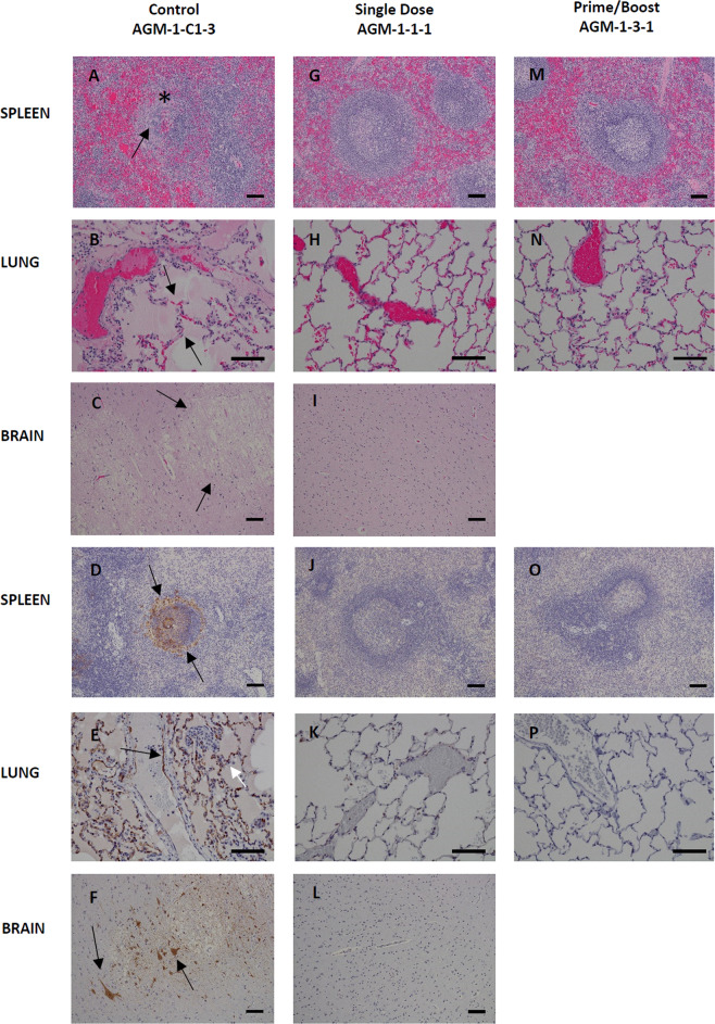
Fig. 5. Histopathology and Immunohistochemistry (IHC) of HeV infected AGM tissues.
Hematoxylin and Eosin (H&E) staining of representative tissues (A–C, G, H, I, M, and N) and HeV antigen staining for IHC images are indicated in brown (D–F, J–L, O, and P). Positive-control AGM for HeV (Control AGM-1-C1-3) includes images A–F. Splenic lymphoid necrosis (*) with syncytial cell formation (arrow) (A) with associated diffuse cytoplasmic immunolabeling of mononuclear cells within the white and red pulp (arrows) (D) interstitial pneumonia (arrows) with alveolar hemorrhage, fibrin, foamy alveolar macrophages, and cellular debris (B) with associated immunolabeling of endothelium (black arrow), alveolar septate (white arrow) and alveolar macrophages (E), diffuse gliosis with vacuolar plaque (arrows) (C) with associated immunolabeling of neuronal cells (arrows) and endothelium of small caliber vessels within the brain (F). No lesions or immunolabeling was noted in representative tissues of Single dose of vaccine (Single dose AGM-1-1-1) images G–L and prime with a boost of vaccine (Prime/Boost AGM-1-3-1) images M–P. H&E and IHC images of the spleen and brain were captured at 10× and lung at 20× magnification. Error bars—100 µm.

