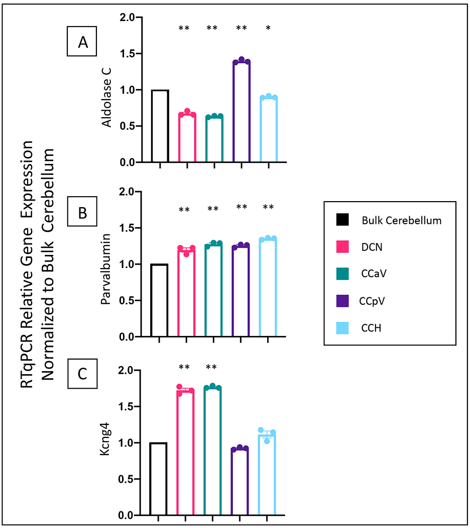
Figure 2: Relative gene expression in isolated specific regions of the cerebellum.

Relative expression of aldolase C (2A), parvalbumin (2B), and Kcng4 (2C) normalized to Rps18 (protein associated with ribosomal RNA, expressed in all cells), and using bulk cerebellar extract as a reference. As expected, aldolase C expression was higher in the posterior vermis and lower in the DCN and anterior vermis (2A). As expected, based on Allen Brain Atlas, parvalbumin expression is uniformly expressed across each extracted region, while Kcng4 expression is significantly enriched in the DCN and CCaV. One-way ANOVA, with a Tukey’s post hoc test. *p<.005, ** p<.0001 relative to the bulk cerebellar extract. Histograms represent average values for N=3 with values for each individual mouse shown as dots. Error bars represent standard error of the mean. DCN (Deep cerebellar nuclei), CCaV (the cerebellar cortex of anterior vermis), CCpV (the cerebellar cortex of the posterior vermis), and CCH (the cerebellar cortex of the hemispheres). N=3 mice for cerebellar dissection regions. N=1 bulk extract. Experiment done in triplicate.
