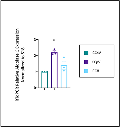
Figure 3: RTqPCR Relative gene expression of aldolase C across cerebellar cortex.

Relative expression of aldolase C in specific regions of the cerebellar cortex – anterior vermis (CCaV), posterior vermis (CCpV), and hemispheres (CCH). Gene expression level of aldolase C was normalized to Rps18 and compared to the expression level in the anterior vermis. As expected aldolase C expression was enriched in the posterior vermis. One-way ANOVA, with a Tukey’s post hoc test. *p<.005 relative to CCaV. Error bars represent standard error of the mean. CCaV (the cerebellar cortex of anterior vermis), CCpV (the cerebellar cortex of the posterior vermis), and CCH (the cerebellar cortex of the hemispheres). N=3 mice, experiment done in triplicate.
