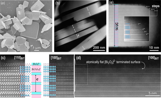
Figure 1.
(a) SEM image of Bi4Ti3O12 template particles. (b) Low-magnification HAADF-STEM image of edge-on-oriented Bi4Ti3O12 platelets. Arrows denote the c axes. (c) Atomic-resolution Z-contrast images of platelets in the [100] Bi4Ti3O12 and [110] Bi4Ti3O12 orientations taken near the surface of the particles with overlaid structural models. The platelets are terminated by the [Bi2O2]2+ layer. (d) Atomically flat (smooth) surface of the platelets as a result of layer-by-layer growth in molten salt. (e) Edge of a platelet with a thickness of 14 unit cells (UCs) (+ an additional [Bi2O2]2+ layer) with steps. The magnified region shows weakly bonded adatoms on the exposed surface on the lateral side of the platelet.

