Figure 4.
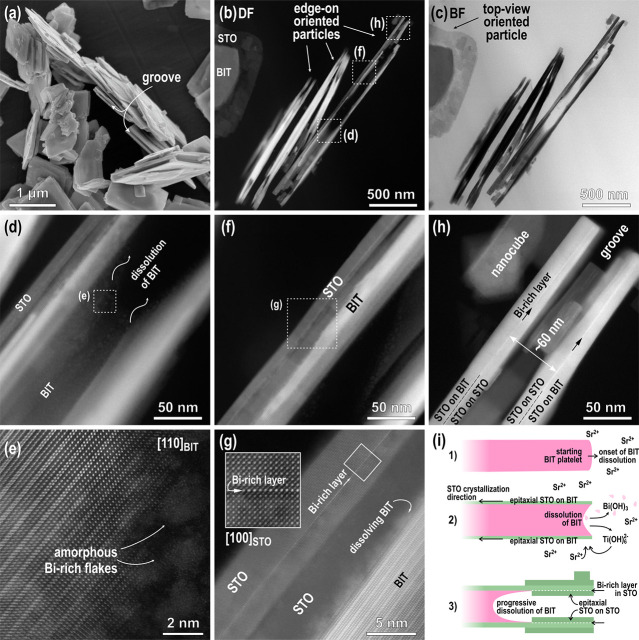
(a) SEM and (b–h) STEM micrographs of SrTiO3/Bi4Ti3O12 heterostructural platelets (mainly edge-on-oriented platelets, which were thinned to electron transparency) as obtained after 1 h of a reaction at 200 °C (6 M NaOH, Sr/Ti = 12). (b) DF and (c) BF images showing (mainly) edge-on platelets along the whole side length; (d) DF of the central part of the platelet (showing SrTiO3 layers and partially disintegrated Bi4Ti3O12 inside the groove). (e) HR image of the area marked in (d) showing the disintegration of Bi4Ti3O12. (f) Another DF of the part between the central area and the edge of the platelet. (g) Magnified area from the image (f) presenting dissolution of Bi4Ti3O12 and as-formed SrTiO3 with the incorporated Bi-rich layer (HR image of this part is in the inset). (h) DF image of the edge of the platelet with two parallel SrTiO3 platelets with incorporated Bi-rich layers. (i) Schematically shown processes of the TC reaction from Bi4Ti3O12 to SrTiO3 as reconstructed from STEM results, presented in (b)–(h).

