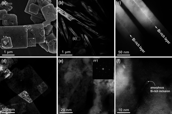
Figure 5.
(a) SEM and (b–f) STEM DF images of (b, c) edge-on oriented SrTiO3 platelets and (d–f) STEM top-view of SrTiO3 platelets, formed after 15 h at 200 °C (Sr:Ti = 12, 6 M NaOH)). (e) STEM image from the central part with FFT showing a (100) orientation in the inset (f) HR image from the central part of the SrTiO3 platelet with Bi-rich inclusion.

