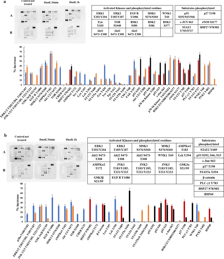
Fig. 4.
Human Phospho-Kinase Arrays for SH-SY5Y not differentiated cells (a) and SH-SY5Y differentiated cells (b) and relative percentage increase of protein phosphorylation after 20 and 60 min of incubation with the exogenous and purified DnaK. The signal produced is proportional to the amount phosphorylation in the bound analyte. The spot signals have been normalized to the internal reference spots first and then to the corresponding not treated sample. The table lists the activated kinases and substrates with the relatives phosphorylated residues with signal increase above 30% of the negative control. In blue: activated Kinase and phosphorylated residues after 20 min of incubation with DnaK; in orange: activated Kinase and phosphorylated residues after 60 min of incubation with DnaK; in red: substrates phosphorylated after 20 min of incubation with DnaK; in black: substrates phosphorylated after 60 min of incubation with DnaK

