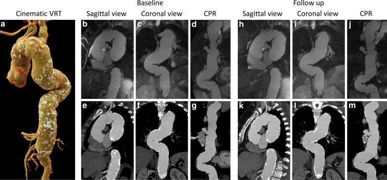
Fig. 5.
Representative case example demonstrating disease monitoring over a 1-year follow up. Cinematic volume rendering technique (a), generated from the baseline CTA of a 46-year-old woman, shows the overview of an extensively calcified and tortuous aorta. Aneurysmal aortic dilatation is present at the level of the root, ascending and descending aorta, extending to the level of the celiac artery. Corresponding CMRA (top row, b–d and h–j) and CTA (bottom row, e–g and k–m) images in sagittal, coronal and curved planar reformat views are shown at baseline (b–g) and at 1-year follow up (h–m). The maximum diameter increased from 47.4 to 53.0 mm (11.8%) in the ascending aorta, and from 53.2 to 54.3 mm (2.1%) in the descending aorta. VRT volume rendering technique, CPR curved planar reformat

