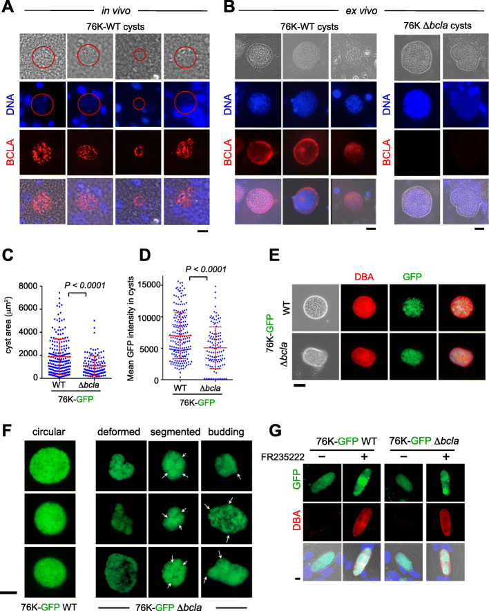
Fig. 5.
BCLA localizes within the cyst matrix and at the cyst periphery while bcla disruption induces subtle changes in cyst morphology. a, b BCLA (red) and DNA (blue) were detected in histological sections of brains (a, in vivo) or in cysts purified from brains (b, ex vivo) in mice chronically infected with 76K-GFP-luc WT or Δbcla strains. Scale bar, 10 μm. c, d Comparative analysis of the area (c) and the GFP-fluorescence intensity (d) of WT and Δbcla-containing cysts purified from the brains of NMRI mice that survive to challenge (in Fig. 4a) showed that loss of bcla translated into smaller cysts with reduced parasite load. e DBA staining (red) of the glycosylated wall from cysts isolated from mice chronically infected with 76K-GFP-luc WT or Δbcla strains. Scale bar, 40 μm. f Representative panels of Δbcla-containing cysts harboring deformations, segmentations, and “buds” compared to the round and well-defined WT cysts. Scale bar, 40 μm. g DBA (red) staining of the glycosylated membrane surrounding in vitro-converted bradyzoites. HFFs were infected with 76K-GFP-luc or 76K-GFP-luc-Δbcla tachyzoites and treated with vehicle (DMSO) or low dose of FR235222 (25 ng/mL for 7 days). Lectin DBA labeling is similar in the membranes surrounding in vitro-converted bradyzoites of Δbcla parasites and the WT strain. Scale bar, 10 μm

