
Fig. 2.
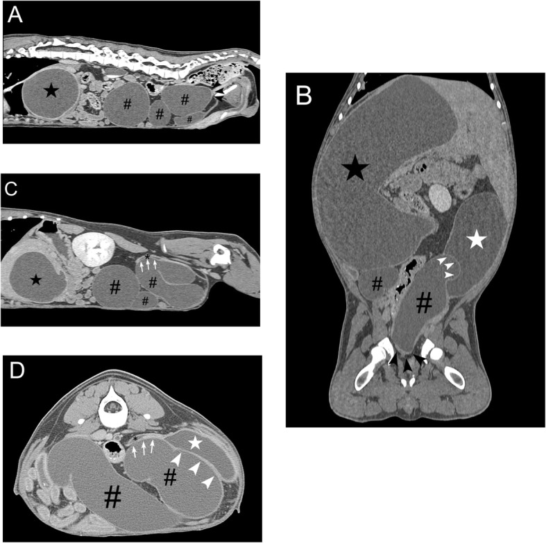
Abdominal computed tomography findings for a dog with distal ureteral atresia. a Sagittal view showing blind-ended termination of the right ureter (black arrowhead). Secondary hydronephrosis (black star) and hydroureter (pound sign) is also present. b Dorsal view showing compression (white arrowhead) of the urinary bladder (white star) by the dilated atresic ureter (pound sign) and hydronephrotic kidney (black star). The blind-ended termination of the right ureter (black arrowhead) is also present. c Sagittal view showing compression (white arrow) of the left distal ureter (black asterisk) by the dilated atresic ureter (pound sign) and hydronephrotic kidney (black star). d Transverse view showing compression (white arrowhead and arrow) of the left distal ureter (black asterisk) and urinary bladder (white star) by the dilated atresic ureter (pound sign)