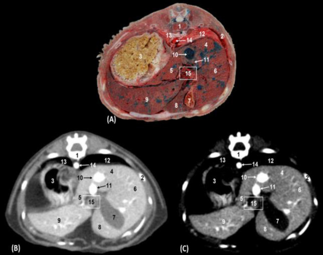
Fig. 2.
Transverse sections of the cranial abdomen at the level of the thirteenth thoracic vertebra (level I). (A) Anatomical section, (B) Soft tissue window CT image, and (C) Mediastinum-vascular window CT image. Caudal views. 1: Thoracic vertebra: body, 2: Rib: body, 3: Stomach: body, 4: Liver: caudate lobe; caudate process, 5: Liver: Caudate lobe; papillary process, 6: Liver: right lateral lobe, 7: In liver gallbladder: quadrate lobe, 8: Liver: left medial lobe, 9: Liver: left lateral lobe, 10: Caudal vena cava, 11: Portal vein (in A bloodless), 12 = Right lung: caudal lobe, 13: Left lung: caudal lobe, 14: Thoracic aorta, and 15: Porta of liver

