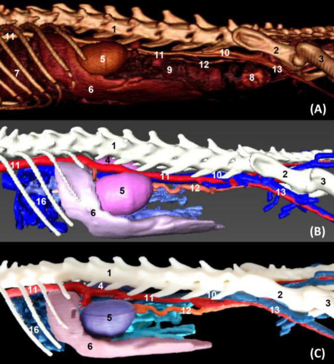
Fig. 7.
Volumetric reconstruction (volume rendering) of the abdominal and pelvic cavities together with the bones of the spine, ribs, and hip joint performed with the (A) OsiriX program, (B) Amira for Fei Systems, and (C) Colored three-dimensional printing. Left side view. 1: Lumbar vertebrae, 2: Ilium: body, 3: Femur, 4: Right kidney, 5: Left kidney, 6: Spleen, 7: Liver, 8: Descending colon, 9: Intestine: coils, 10: Caudal vena cava, 11: Abdominal aorta, 12: Left testicular artery, and 13: External iliac artery and vein

