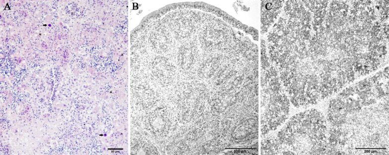
Fig. 2.
Histopathology of the affected chicken associated with FAdV-11. (A) Area of necrosis appears with hepatic cords disruption, eosinophilic cellular remainders, and inflammatory cells infiltration. Typical large, round, deeply basophilic intranuclear inclusion bodies are present in the necrotic area (arrows) (H&E, scale bar = 50 μm), (B) Mild loss of the follicular lymphoid cell population of the cloacal bursa (H&E, scale bar = 200 μm), and (C) Mild depletion of the lymphocytes in both the cortexes and medullae of the thymus (H&E, scale bar = 200 μm)

