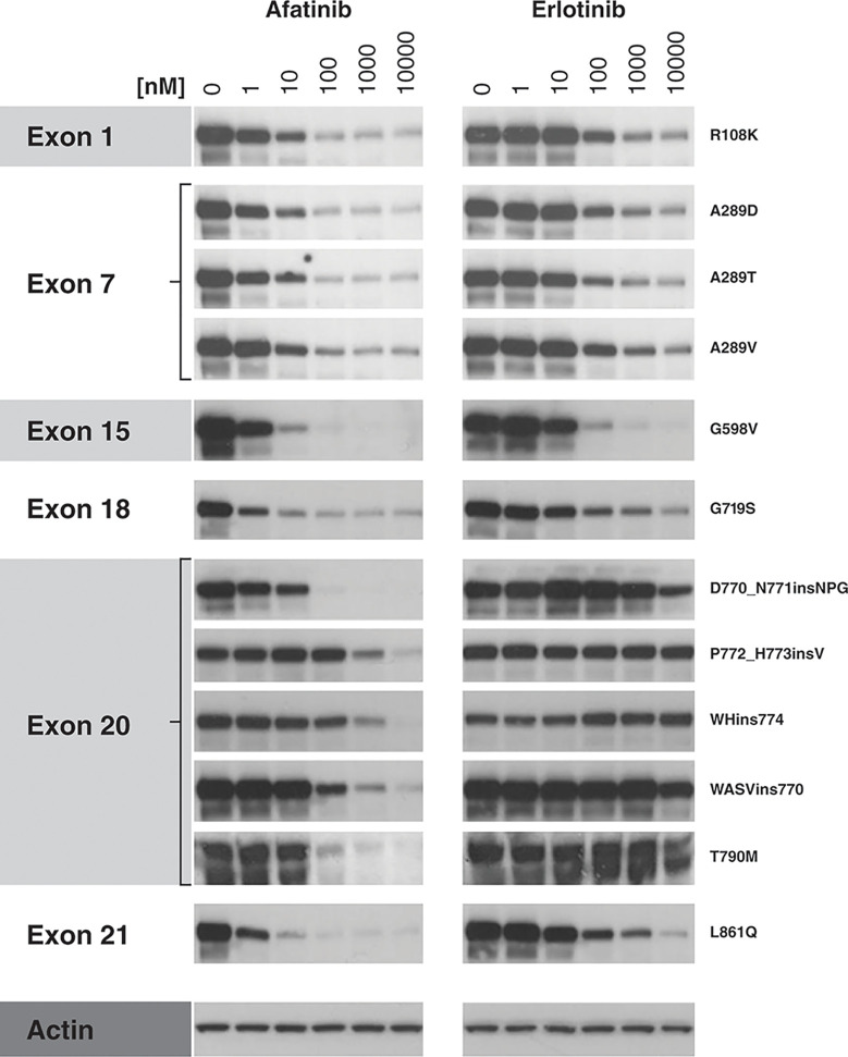
Figure 1.
Inhibition of EGFR mutant protein autophosphorylation by afatinib and erlotinib in cellular assays. NIH-3T3 cells (ATCC; #CRL-1658) were cultured in supplemented Dulbecco’s Modified Eagle Medium and cultivated at 37°C/5% CO2 in a humidified atmosphere to maintain <80% confluence. Cells were then transfected with one of 12 EGFR mutant plasmid constructs, using 4 µg of DNA for EGFR variants G598V, D770_N771insNPG, P772_H773insV, WHins774, and T790M, and 2.5 µg of constructs R108K, A289D, A289T, A289V, G719S, WASVins770, and L861Q, diluted in 250 µl serum-free culture medium. A diluted Lipofectamine-DNA mix was then added drop-wise to the cells. 48 h post-transfection, cells were treated for 2 h with afatinib or erlotinib (1–10,000 nM) or were left untreated. At 2 h post-treatment, protein lysates were prepared using lysis buffer and the effect of TKI treatment on EGFR tyrosine-1068 phosphorylation was analyzed by Western blot (primary antibody: phospho-specific anti-EGFR [Y1068] antibody; Abcam, #ab40815, 1:1,000 dilution; secondary antibody: goat anti-rabbit; Dako, #P0448, 1:1,000 dilution). Actin was used as a loading control. EGFR, epidermal growth factor receptor.

