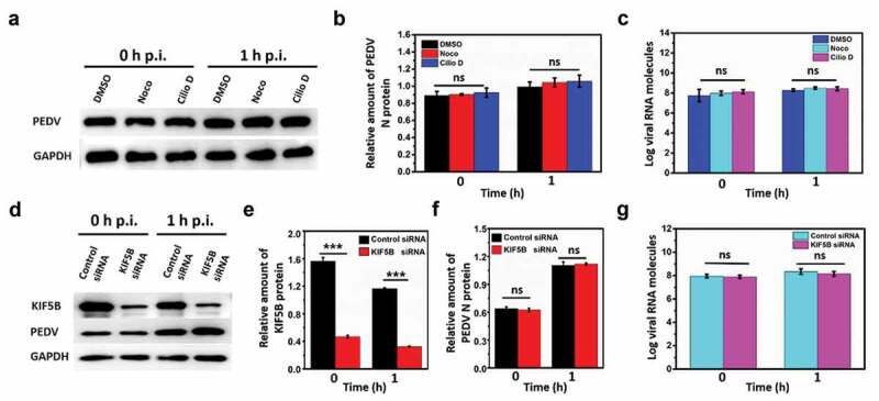
Figure 3.

Microtubule, dynein and kinesin-1 are not required for PEDV attachment or internalization. (a) Detection on PEDV N protein and GAPDH using Western blot from the infected PEDV particles in the control DMSO, Noco and Cilio D pretreated Vero cells at 0 h and 1 h PEDV post-infection (p.i.). (b) Quantification on the amount of PEDV N protein according to the amount of GAPDH. (c) Copy numbers of PEDV RNA from the infected PEDV particles in the control DMSO, Noco and Cilio D pretreated Vero cells at 0 h and 1 h PEDV post-infection using RT-PCR. (d) Detection on the proteins of KIF5B, PEDV N protein and GAPDH using Western blot from the infected PEDV particles in the control siRNA and KIF5B siRNA pretreated Vero cells at 0 h and 1 h PEDV post-infection (p.i.). (e) and (f) Quantification on the amount of KIF5B and PEDV N protein according to the amount of GAPDH. (g) Copy numbers of PEDV RNA from infected PEDV particles in the control siRNA and KIF5B siRNA pretreated Vero cells at 0 h and 1 h PEDV post-infection using RT-PCR. Each data point represents mean ± standard deviation from three independent experiments. Statistical analysis of all data was performed using one-way ANOVA (***, P < 0.001). ns: nonsignificant
