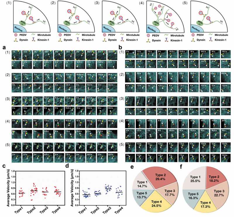
Figure 4.

Five types of PEDV intercellular transport observed by single-virus tracking in live Vero cells. (a)-(b) Representative time-lapse fluorescence images of PEDV particles during intercellular transport in live Vero cells transfected with (a) EGFP-MT and mKO2-dynein and (b) EGFP-MT and mKO2-KIF5B in different types: (1) unidirectional movement toward microtubule plus ends, (2) unidirectional movement toward microtubule minus ends, (3) bidirectional movement along the same microtubule, (4) bidirectional movement along different microtubules and (5) motionless state. White arrows indicate PEDV particles. Scale bar, 1 μm. (c) Average velocities corresponding to motion states of PEDV intercellular transport according to (a). (d) Average velocities corresponding to motion states of PEDV intercellular transport according to (b). (e) Proportions corresponding to five types of PEDV intercellular transport according to (a). (f) Proportions corresponding to five types of PEDV intercellular transport according to (b)
