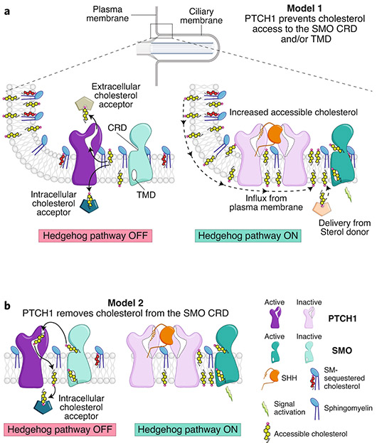
Fig. 7 ∣. Two models for the regulation of SMO by PTCH1 at primary cilia.
a, A cartoon representation of cilia is shown at the top, with a rectangle showing a region at the cilia base known as the ‘ciliary pocket’, magnified below. In model 1, PTCH1 uses its transporter function (left) to remove cholesterol from the ciliary membrane, transferring it to an acceptor. Because of the higher abundance of SM in the ciliary membrane, accessible cholesterol drops below the threshold required to activate SMO. Inactivation of PTCH1 (right) allows influx of cholesterol back into the ciliary membrane, driving SMO activity. b, An alternative model in which PTCH1 directly inactivates SMO by accepting cholesterol from its CRD, inspired by how cholesterol bound to the NTD of NPC1 is transferred to the CTD (see Fig. 3b).

