
FIGURE.
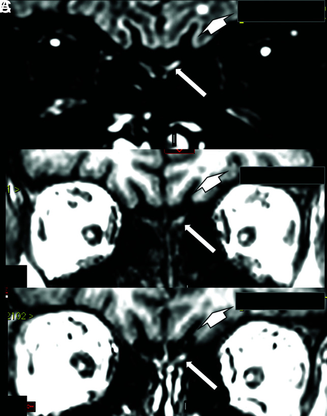
A woman in her 60s with confirmed COVID-19 for which MR imaging shows a hyperintense lesion on a coronal reconstruction of 3D-FLAIR (A, arrow) and also on pre- (B, arrow) and postcontrast SPGR T1WI (C, arrow). This is suggestive of a component of probably methemoglobin in this left olfactory bulb lesion, which seems to be a little larger and asymmetric compared with the apparently normal right olfactory bulb. This asymmetry is better seen on FLAIR. On FLAIR (A, short arrow), there is also a small round hyperintense lesion in the subcortical white matter in the left frontal lobe, which is hypointense on T1WI (B, short arrow) and does not enhance on postcontrast T1WI (C, short arrow), being no specific lesion area probably corresponding to gliosis. This patient also had some areas of brain parenchymal bleeding and microbleeding, and had around 50% of the bilateral pulmonary parenchyma compromised with typical COVID-19 lesions on chest CT, not shown in this figure.