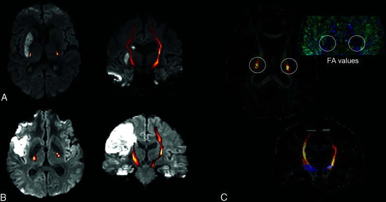
FIG 2.
Examples of 2 patients 3 days after mechanical recanalization of a right middle cerebral artery occlusion and consecutive basal ganglia infarction (A, patient 1)/peripheral infarct (B, patient 2). The CST, reconstructed by probabilistic fiber tracking (C, demonstrated on FA maps, blue seed ROI, gray target ROI, yellow posterior limb of the internal capsule) is overlaid on the reconstructed DWI trace picture in A and B (CST connectivity values, increasing probability from red to yellow before CST thresholding). For both patients, lower FA values of the CST within the posterior limb of the internal capsule are found for the infarcted right side in comparison with the healthy, nonaffected left side (C, illustrated using 3D Slicer; http://www.slicer.org).

