
Extended Data Figure 9. P3 RO saline- and ABE AAV9-injected male and female mouse skin histology assessed by H&E and Masson staining.
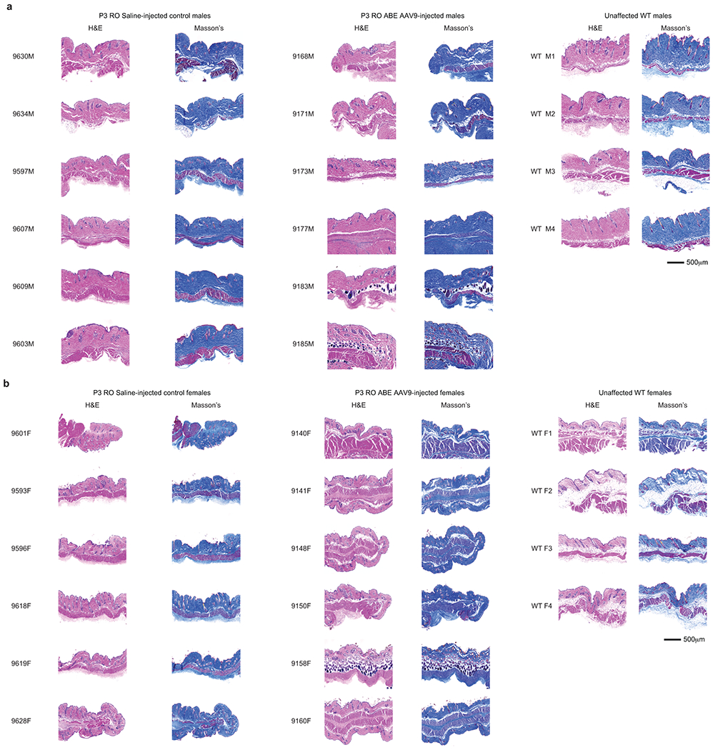
(a) Representative skin cross-sections for P3 RO saline-injected (left), ABE-AAV9-injected (middle), and wild-type C57BL/6 males at 6 months of age. Left images were stained with hematoxylin and eosin (H&E); right images were stained with Masson stain. (b) Representative skin cross-sections for P3 RO saline-injected (left), ABE-AAV9-injected (middle), and wild-type C57BL/6 females at 6 months of age. Left images were stained with hematoxylin and eosin (H&E); right images were stained with Masson stain. Unaffected WT are wild-type C57BL/6 mice. These sections each represent replicates from different mice.