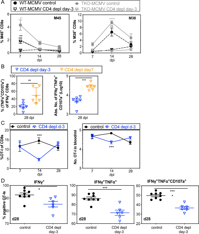
Fig 5. CD4+ T cell help is needed for the proliferation and function of MCMV-specific CD8+ T cells.
A. CD8+ T cells are reduced in frequency after i.n. infection in the absence of CD4+ T cell help. Shown is the frequency of viral tetramer-specific CD8+ T cells in the blood of recipients at the indicated time points with or without CD4+ T cell depletion before infection. Data show the average frequency of T cells at day 7 (n = 9–12), day 14 (n = 6–9) and day 28 (n = 3–6) after i.n. infection and are derived from one representative experiment of at least 3 independent experiments. B. CD8+ T cell function is impaired in the absence of CD4+ T cell help after i.n. infection, but improved by delaying CD4+ T cell depletion until day 7. Each symbol represents an individual animal. The solid line shows the mean value, and error bars represent the SEM. Data are combined from two independent experiments. C. The frequency and number of OT-Is are impaired in the absence of CD4+ T cell help after i.n. infection with MCMV-Ova. CD4+ T cells were depleted or not from C57BL/6 mice. One day before i.n. infection with MCMV-Ova, mice received 5000 OT-I T cells. Shown are the frequency (left) and absolute number (right) of OT-I cells in blood over time after infection. Data show the average values from 6–8 animals per group, and error bars represent the SEM. CountBright absolute counting beads (ThermoFisher Scientific) were included to determine the number of cells in the blood. D. The function of OT-I T cells in the spleen is impaired on a per-cell basis in the absence of CD4+ T cell help. OT-I T cells in the spleens of adoptive recipients (as described in C.) were analysed for production of IFN-γ, TNF-α and degranulation (exposure of CD107a) after stimulation with SIINFEKL peptide 28 days after infection. Each symbol represents an individual animal. The solid line shows the mean value, and error bars represent the SEM.

