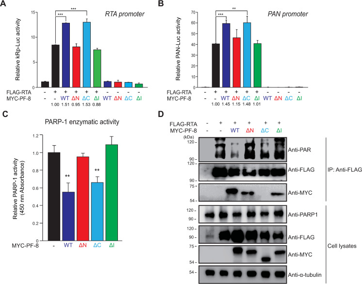
Fig 4. Binding of PARP1 to PF-8, not RTA, is essential for PF-8–induced RTA transactivation enhancement.
The latter is mediated by decreased levels of poly(ADP-ribosyl)ated (PARylated) RTA. (A and B) Luciferase reporter assays of PF-8 mutants. HEK293T cells were transfected with reporter construct pGL3-kRP-Luc (A) or pGL3-PAN-Luc (B) (300 ng) and MYC-PF-8 mutants (150 ng) in the presence or absence of the FLAG-tagged RTA expression plasmid (25 ng). The cells were harvested at 48 h post-transfection for luciferase reporter assays. Each transfection was performed in triplicate, and the EGFP-expressing plasmid served as an internal control. The increased fold values of promoter activity relative to the RTA alone sample are indicated. Statistical analysis was performed by Student’s t test (**P < 0.01 and ***P < 0.005). (C) PARP1 activity in the cells expressing PF-8 mutants. HEK293T cells were transfected with MYC-tagged PF-8 mutants. The transfected cells were harvested at 48 h post-transfection. The PARP1 inhibition activity in 50 μg of cell lysates was analyzed using the PARP1 assay kit with histone-coated strip wells at 450 nm absorbance. Statistical analysis was performed by Student’s t test (**P < 0.01). (D) PF-8 mutant–mediated PARylation of RTA. HEK293T cells were transfected with MYC-tagged PF-8 mutants and FLAG-tagged RTA constructs. The transfected cells were harvested at 48 h post-transfection and subjected to an immunoprecipitation assay with the anti-FLAG-M2 antibody. The cell lysates were investigated by western blotting with the anti-PAR, anti-PARP1, anti-FLAG-M2 anti-MYC, and anti-α-tubulin antibodies.

