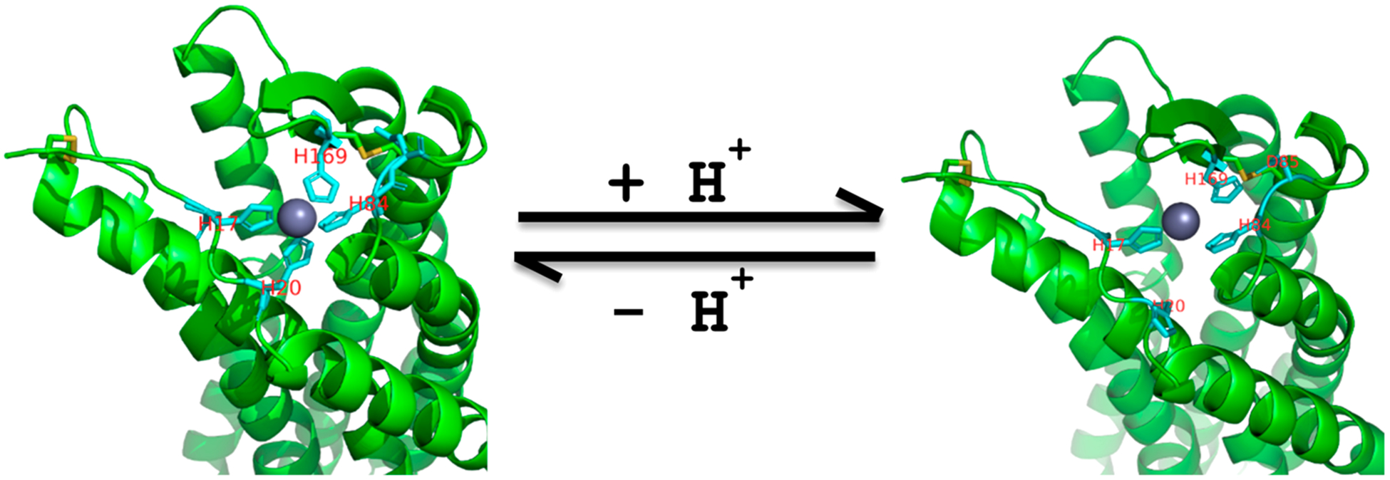
Figure 11.

Extracellular divalent metal ion binding models. Left, H17, H20, H84, and H169 of GPR68 form tetradentate coordination with Zn2+ under high pH conditions. Right, protonated H20 breaks away, and protonated H169 breaks away and forms interactions with nearby D85. Two disulfide bonds (in yellow) help constrain the Zn2+ binding site.
