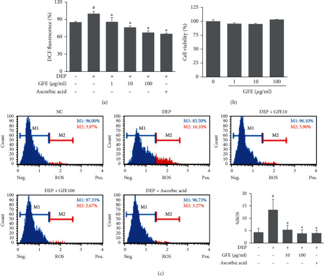
Figure 2.

Effects of GFE on ROS production in DEP-stimulated HaCaT cells. (a) Percentages of cells exhibiting oxidative stress (ROS positive cells) were determined using an Oxidative Stress Kit. Cells were pretreated with GFE (10, 100 μg/ml) for 1 h and then stimulated with DEP (100 μg/ml) for 12 h. (b) Cytotoxic effects of GFE in HaCaT cells. Cells were treated with GFE (1, 10, 100 μg/ml) for 24 h and cell viabilities were assessed using an XTT kit. Data are presented as mean ± SD (n = 3). #P < 0.05 vs. normal controls (NC), ∗P < 0.05 vs. DEP treated cells.
