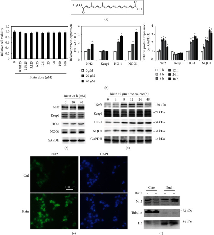
Figure 1.

Bixin activated the Nrf2 signals in liver cells without detectable toxicity. (a) Bixin's chemical structure. (b) Cell viability was measured in LO2 cells administrated with the indicated doses of bixin for 48 h. (c) LO2 cells were administrated with bixin (0-40 μM) for 24 h or treated with bixin 40 μM for the indicated time (d). Cell lysates were subjected to immunoblot analyses with the indicated antibodies. Quantification of relative protein expression was determined; results are expressed as the means ± SD (∗p < 0.05, Ctrl vs. bixin treatments). (e) After treated with bixin (40 μM) for 24 h, LO2 cells were fixed and subjected to indirect immunofluorescence staining of Nrf2 (green); nucleus was stained with DAPI (the representative images were shown, scale bar = 100 μm). (f) Immunoblot analysis of Nrf2 in nuclear and cytoplasmic fractions of LO2 cells with or without bixin treatment with the indicated antibodies. Cyto: cytoplasmic fraction of LO2 cells; Nucl: nuclear fraction of LO2 cell.
