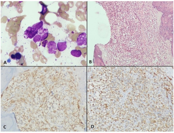
Figure 3.
(A) Bone marrow (BM) aspirate showing normal marrow cells along with degenerated mast cell (MC) (Giemsa and MGG stain ×1000). (B) BM biopsy showing fibrosis and nodular aggregates of clear looking MCs (H&E stain ×200). (C) MCs showing positivity for MC tryptase (MCT stain ×200) (D): MCs showing positivity for CD117 (CD117 stain ×200).

