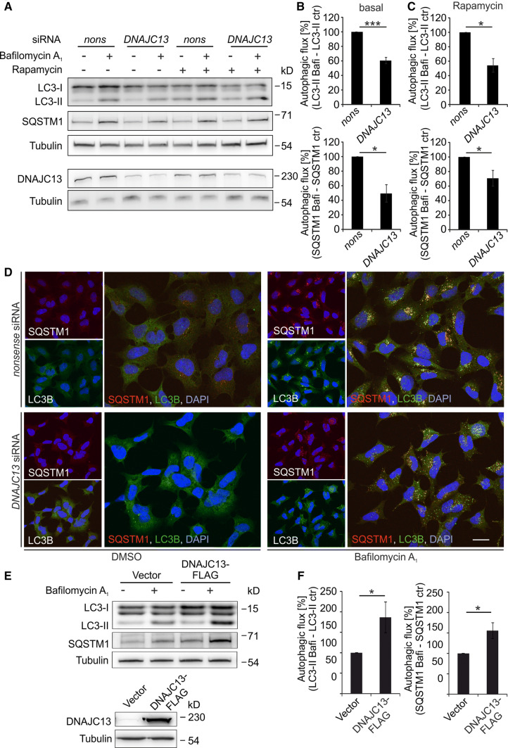
Fig. 2.
DNAJC13 positively modulates autophagy. a–d HEK293A cells were transfected with nonsense (nons) or DNAJC13 siRNA. 48 h after transfection cells were treated with bafilomycin A1 and/or rapamycin for additional 4 h. a–c Cell extracts were separated on SDS-PAGE and transferred on nitrocellulose membrane. Expression levels of LC3B-II and SQSTM1 were quantified and normalized to tubulin. The lower band of the LC3B-I duplet in some blots may represent a processing intermediate [71]. Autophagic flux under basal conditions (b) or rapamycin treatment (c) was determined by subtraction of LC3B-II and SQSTM1 levels without bafilomycin A1 (ctr) from LC3-II and SQSTM1 levels with bafilomycin A1 (Bafi), respectively (n = 4 [basal], n = 3 [rapamycin]; mean ± SEM; t test: *p ≤ 0.05, ***p ≤ 0.001). d Confocal images of HEK293A cells transfected with nonsense or DNAJC13 siRNA as above and stained for SQSTM1 and LC3B. The nucleus is detected by DAPI (scale bar: 20 µm). e, f Autophagic flux in HEK293T cells transiently overexpressing DNAJC13-FLAG was determined as in a, b (n = 4; mean ± SEM; t test: *p ≤ 0.05)

