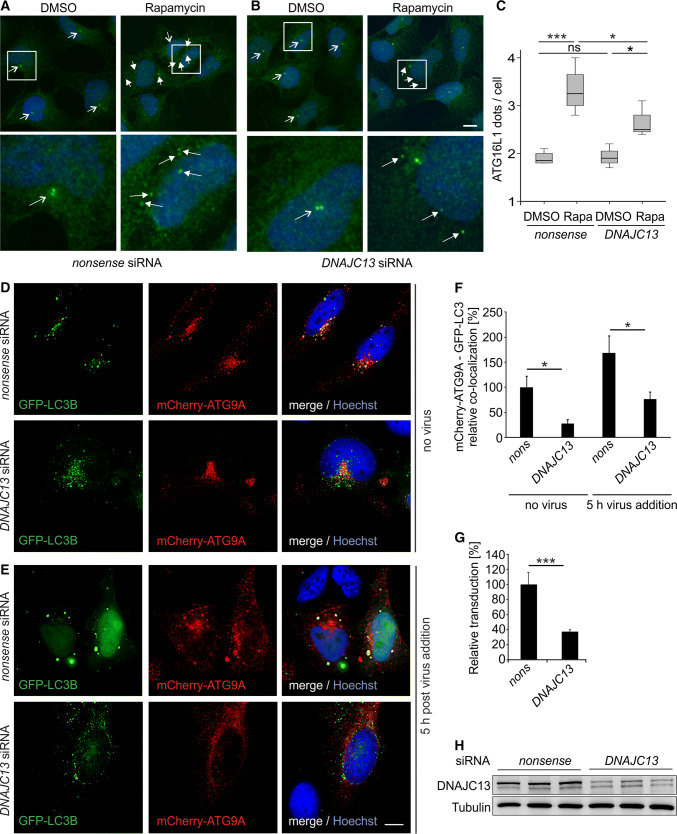
Fig. 4.
DNAJC13 affects the formation of ATG16L1 puncta and the co-localization of ATG9A with LC3. HEK293A cells transfected with a nonsense or b DNAJC13 siRNA and treated with rapamycin were stained with antibodies detecting ATG16L1 (green) and DAPI (blue). Images represent maximum projections of image stacks. The lower panels are enlargements of the boxed areas. All cells carrying a single or a double point in close proximity to the nucleus (open arrows) represent cells with a low autophagic activity; multiple, accumulated ATG16L1 dots (closed arrows) reflect a high autophagic activity (scale bar: 10 µm). c In average, dots in 60 cells per condition and experiment were counted (n = 4; box-plot; one-way ANOVA with Bonferroni correction: *p ≤ 0.05; ***p ≤ 0.001). d, e HeLa cells were transfected with nonsense (nons) or siRNA against DNAJC13 and GFP-LC3B (in green) and mCherry-ATG9A (in red). Representative deconvoluted images of z-stacks are shown without infection (d) and 5 h post-HPV pseudovirion addition (e). DNA was labeled with Hoechst and is shown in blue (scale bar: 10 µm). f Analysis of GFP-LC3B and mCherry-ATG9A resulted in a decreased co-localization of pixels upon knockdown of DNAJC13. The infection with HPV virions resulted in an increased co-localization of ATG9A and LC3B and the knockdown of DNAJC13 affects this co-localization. About eight images per condition were analyzed in three independent experiments each (about 100 cells in total). Values were normalized to nonsense siRNA transfected cells without virus (n = 24; mean ± SEM; Wilcoxon rank-sum test: *p ≤ 0.05). g HeLa cells treated with nonsense or DNAJC13 siRNA were infected with HPV virions containing a luciferase expression plasmid. Only cells in which the plasmid reached the nucleus expressed luciferase. Relative infection was measured by luciferase activity and normalized by LDH measurements. Control siRNA infection rate was set to 100% (n = 4; mean ± SD; t test: ***p ≤ 0.001). h DNAJC13 protein levels were determined after transfection of HeLa cells with nonsense or DNAJC13 siRNA by Western blot. Tubulin served as a loading control

