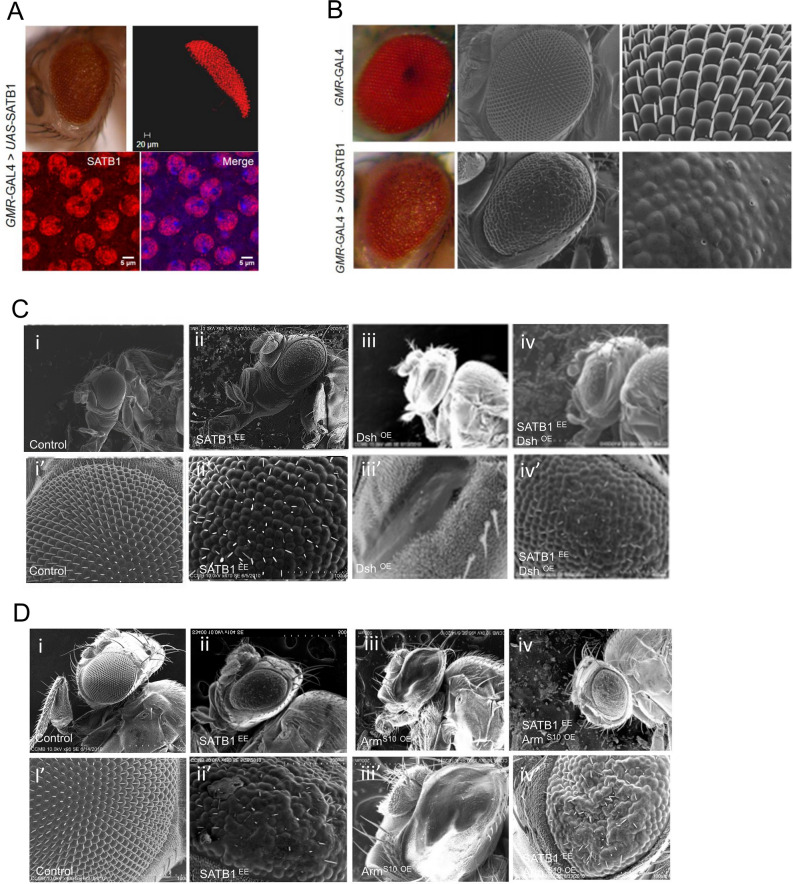
Figure 3.
Subcellular localization of SATB1 is comparable in mammalian cell lines and cells of the eye imaginal discs. (A) Ectopic expression of human SATB1 in the fly eye gives rise to rough eye. Immunostaining for ectopically-expressed SATB1 (red) and DNA (blue) in the eye imaginal disc revealed that SATB1 is localized in the region posterior to the morphogenetic furrow (top panel). Higher magnification immunofluorescence images reveal that similar to that in the mammalian system, SATB1 localizes entirely within the nucleus, while completely excluding the heterochromatic regions. (B) Scanning electron microscopic (SEM) images depicting the fusion of ommatidia as a result of ectopic expression of full-length SATB1. SEM imaging of GMR-Gal4 > UAS-SATB1 fly eyes revealed that ommatidia have lost their wild type hexagonal shape and the compact arrangement. Inter-ommatidial bristles are randomly arranged while some regions of the eye completely lack bristles (Penetrance 100%, n = 110). (C) Small-eye phenotype due to Dsh overexpression is suppressed by expression of human SATB1. (i) Control GMR-GAL4/+. (ii) Fly eyes ectopically expressing SATB1. (iii) Fly eyes ectopically expressing Dsh have small eyes. (iv) Fly eyes expressing SATB1 in the background of Dsh misexpression have large eyes as compared to GMR-Gal4 > UAS-dsh (Penetrance of this phenotype is 30%, n = 200) (D) Small-eye phenotype due to expression of activated armS10 is suppressed by expression of human SATB1. (i) Control GMR-GAL4/+. (ii) Fly eyes ectopically expressing SATB1. (iii) Fly eyes ectopically expressing ArmS10 exhibit the small-eye phenotype. (iv) Flies expressing SATB1 in the background of ArmS10 expression have large eyes as compared to GMR-Gal4 > UAS-armS10 (Penetrance of this phenotype is 20%, n = 174) (I”), (ii’), (iii’), and (iv’) in C and D are higher magnification images. At all stages UAS-GFP was used as control.

