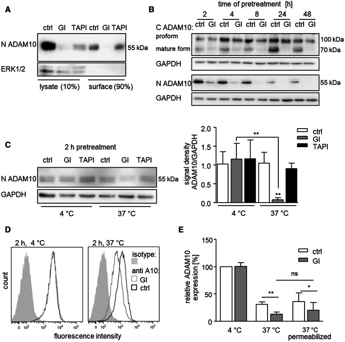
Fig. 2.
Effect of GI on ADAM10 in surface precipitates and cell lysates and temperature dependence of ADAM10 downregulation. a THP-1 cells were treated with 10 μM GI, 10 μM TAPI or vehicle control for 4 h. Surface proteins were biotinylated on intact cells and subsequently precipitated from cell lysates. Precipitates and lysates were then probed by western blotting with an antibody against the N-terminus of ADAM10. Detection of total cytosolic ERK1/2 served as a control. b THP-1 cells were treated with 10 μM GI or vehicle control for the indicated periods of time. Cells were lysed and lysates were then probed by western blotting with antibodies against the N-terminus or C-terminus of ADAM10 or against GAPDH as loading control. c THP-1 cells were treated with 10 μM GI, 10 μM TAPI or vehicle control for 2 h at either 4 °C or 37 °C. Cells were cooled on ice, lysed, and probed by western blotting with antibodies against the N-terminus of ADAM10 or against GAPDH as loading control. Data are shown as representative western blot and as relative changes of band intensity determined by densitometric analysis. a–c One representative western blot out of three independent experiments. d–e THP-1 cells were first stained with primary antibody against ADAM10, then treated with 10 μM GI or vehicle control for 2 h at either 4 °C or 37 °C and afterwards incubated with the secondary antibody on ice. Cells were either left intact or permeabilized before detection of ADAM10 by flow cytometry. Data were obtained in five independent experiments and are shown as representative histogram (d) or as means and SD of the geometric mean fluorescence in relation to that of the 4 °C control (e)

