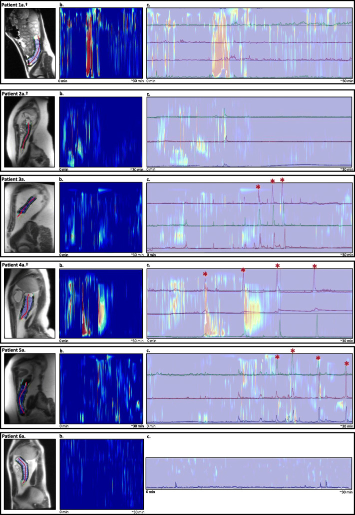
Fig. 2.
Simultaneous colonic manometry and cine magnetic resonance imaging (cine-MRI) motility results. The figure displays: (a) a sagittal view of the child showing the region of interest in the descending colon; (b) the results of about 30 min of cine-MRI colonic activity. Recording time in minutes is displayed on the x-axis. Colonic activity is reported on the y-axis, with red colours representing a small diameter (contraction) and blue colours no change in diameter; and (c) the about 30 min of colonic manometry recordings with an overlay of simultaneous cine-MRI results as shown in b, to allow for a visual comparison of high-amplitude propagating contractions (HAPCs). The x-axis displays the time from the start of simultaneous cine-MRI imaging (0 min) to the termination of the cine-MRI recordings (about 30 min). The y-axis displays the manometric channels in the descending colon with pressure changes (in mmHg), overlapping the cine-MRI motility results as shown in b. ± Patient with caecostomy in place, * HAPC

