Figure 8.
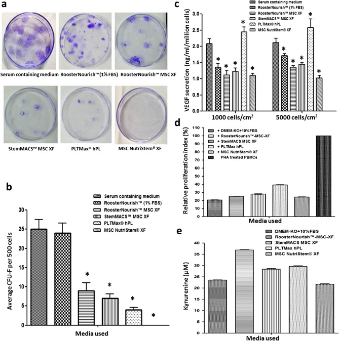
Colony forming ability, potency and immunosuppressive potential of BM-MSCs cultured in SFM/XFM and control medium. (a) Representative photograph of CFU-F of BM-MSCs grown in low serum/ SFM/XFM and control medium. (b) Bar graph showing the number of colonies of BM-MSCs cultured in low serum/SFM/XFM and control medium. The data is represented as mean ± SD (n = 2; *p < 0.05). (c) Cryopreserved cells were seeded into T-75 flasks post revival and grown in respective media for 72 h and the amount of VEGF secreted was determined by ELISA. Results are expressed in concentration (ng/ml/million cells) as mean of duplicate experiment. The amount of VEGF secretion was higher in BM-MSCs cultured in PLTMax hPL when compared to other media and control medium. (d) Bar graph shows the percentage proliferation of PHA stimulated T cell blasts in MSC: PHA activated PBMC co-cultures cultured in SFM/XFM and control medium. The proliferation index in co-cultures is relative to proliferation in PHA blasts (cultured in the absence of MSCs) which was considered as 100% (e) To determine IDO enzyme activity, the culture supernatant of MSC: PHA activated PBMC co-cultures of different media groups was assayed by spectrophotometric detection of the kynurenine concentration (tryptophan metabolite which is a product of IDO catabolism) and is represented as Kynuerurine units (μM). IDO secretion was higher in co-cultures cultured in SFM/XFM when compared to serum-containing medium.

