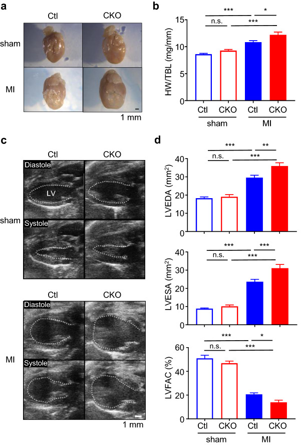
Figure 2.
Deletion of Cxcr7 in cardiomyocytes exacerbates post-infarction remodeling. (a) Representative images of infarcted hearts from Ctl and CKO mice 4 weeks after ligation of a left anterior descending artery to induce myocardial infarction. (b) Heart weight-to-tibia length ratio (HW/TBL) of infarcted mice. sham-Ctl, n = 10; sham-CKO, n = 9; MI-Ctl, n = 15; MI-CKO, n = 9. Data are shown as the mean ± SEM. Significance was calculated by one-way ANOVA followed by the Bonferroni procedure. *P < 0.05, ***P < 0.001. (c) Representative B-mode images of transthoracic echocardiography at diastole and systole of sham and infarcted hearts in Ctl and CKO mice. The dashed line indicates the endocardial surface of the left ventricular (LV) cavity. (d) Left ventricular end-diastolic area (LVEDA), end-systolic area (LVESA), and fractional area change (LVFAC) assessed by echocardiography 4 weeks after myocardial infarction. (sham-Ctl, n = 10; sham-CKO, n = 9; MI-Ctl, n = 15; MI-CKO, n = 9). Data are the mean ± SEM. Significance was calculated by one-way ANOVA followed by the Bonferroni procedure. *P < 0.05, **P < 0.01, ***P < 0.001.

