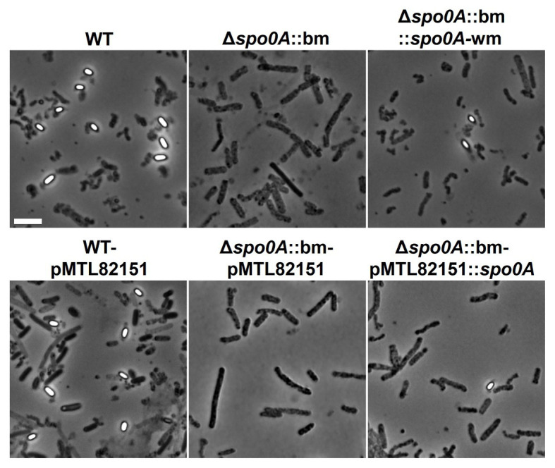
Figure 5.

Phase-contrast microscopy images at 14days post inoculation demonstrating the morphological differences between WT, Δspo0A::bm, and complemented mutant strains. Phase-bright endospores were detectable only in samples containing WT or complemented strains. The Δspo0A::bm mutant did not display any spores or sporulating cells, suggesting that endospore formation ceased at the early stage. Scale bar 5μm.
