


Figure 2.
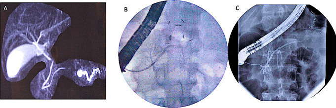
Disconnected pancreatic duct images (A): MRCP showing non-projection of main pancreatic duct in body region with dilated upstream pancreatic duct suggestive of DPD, (B): ERCP showing normal diameter pancreatic duct in head, with cut-off at genu and non-visualisation of upstream duct suggestive of DPD, (C): ERCP showing normal diameter pancreatic duct in head and proximal body with cut-off at distal body and non visualisation of upstream duct suggestive of DPD. DPD, disconnected pancreatic duct; ERCP, endoscopic retrograde cholangiopancreatography; MRCP, MR cholangiopancreatography.