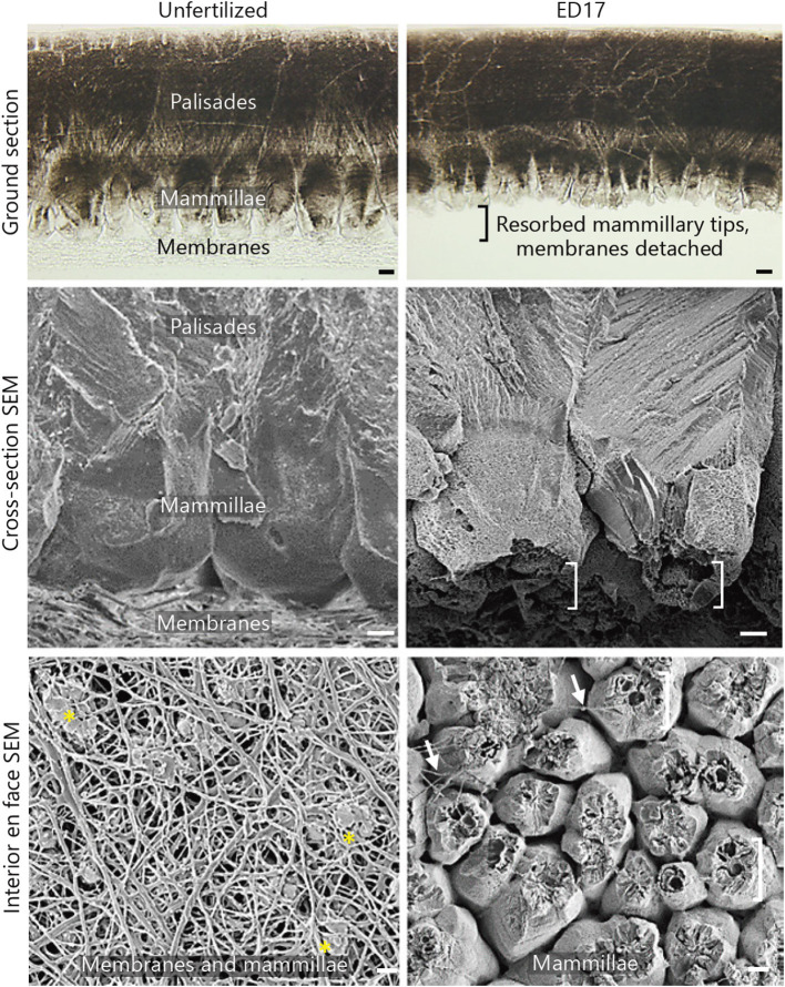
Figure 1.
Ground and polished sections and scanning electron microscopy (SEM) images of the eggshell (ES) and its membranes in unfertilized (left) and fertilized egg (right), showing dissolution of the innermost portion of the ES and detachment of the membranes during incubation of the fertilized egg. Brackets- dissolved mammillary tips; asterisks- intact mammillary tips; arrows- residual membrane fibers. ED17 stands for day 17 of incubation. Scale bars, 10 μm (Hincke et al., 2019).

