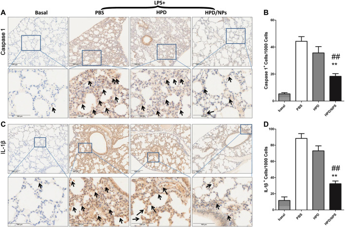
FIGURE 6.
Impact of HPD and HPD/NPs on markers of pyroptosis in lungs of LPS-challenged mice. At 3 h post-LPS, PBS (vehicle), HPD, or HPD/NPs were nasally administered to mice. Lung tissues were collected at 24 h post-LPS challenge. (A) Representative images and (B) quantification of lung tissue cross-sections immune-stained for caspase 1. (C) Representative images and (D) quantification of lung tissue cross-sections immune-stained for IL-1β. Arrows indicate brown/positive staining. N = 4/group; **p < 0.01 vs. PBS, ## p < 0.01 vs. HPD.

