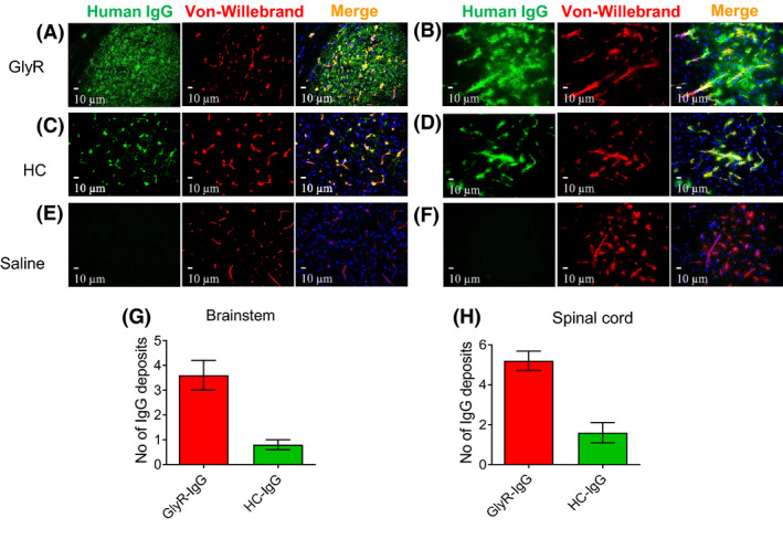
Figure 3.

IgG deposits were found in different brain regions involved in motor control particularly the brainstem and spinal cord. Double labelled photographs were taken at 40× magnification at the brainstem in the PnC and the ventral horn of the spinal cord. In GlyR‐IgG‐treated animals, human IgG (green) is located inside the vessels colocalizing with Von‐Willebrand factor antibody (red), and also in patches within these brain regions without any colocalization (a, b). In healthy control‐treated animals, the human IgG (green) is located only in the vessels (c, d). In saline‐treated animals (e, f) and uninjected controls (not shown), human IgG was not detectable. (g, h) Quantitative analysis shows the mean number of patches of IgG seen in the two IgG‐injected groups, in the brainstem (Two‐sided student t‐tests: t = 4.427 df = 8, P = 0.002) (g) and in the spinal cord (t = 5.091 df = 8, P < 0.001) (h). Nuclei are stained with DAPI (blue).
