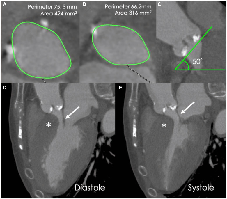
Figure 2.
Pre-procedural cardiac computed tomography images. (A) Annular assessment. (B) Left ventricular outflow tract assessment. (C) The angulation of the aorta was 50°. (D, E) Long-axis computed tomography images of the diastolic and systolic phases showing interventricular septal bulge (*). Asymmetric septal hypertrophy or systolic anterior motion of the anterior mitral leaflet are not observed. Anterior mitral leaflet (arrow).

