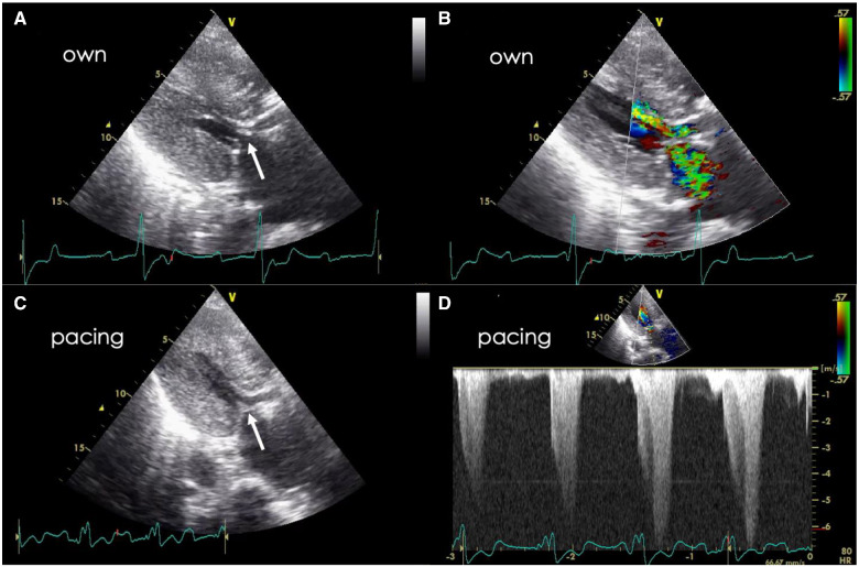
Figure 4.
Transthoracic echocardiography after transcatheter aortic valve implantation. (A, B) Under own junctional rhythm. Left ventricular outflow tract obstruction with systolic anterior motion of the mitral valve (arrow). The anterior mitral leaflet is thickened. (C) Systolic anterior motion of the anterior mitral leaflet (arrow) and left ventricular outflow tract obstruction are decreased by right ventricular pacing. (D) The peak flow velocity and pressure gradient are 6.8 m/s and 185 mmHg, respectively, even under right ventricular pacing.

