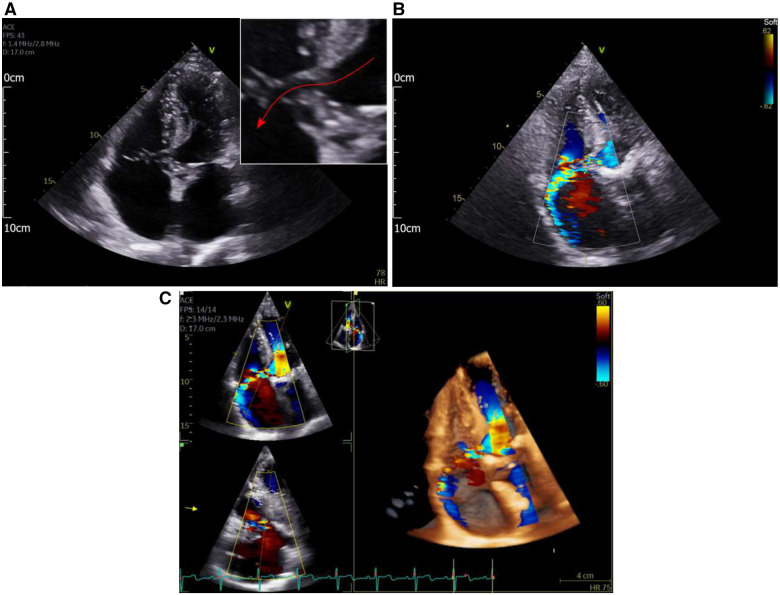
Figure 1.
(A) Apical four-chamber view demonstrating a hypoechoic tract between the left ventricular outflow tract and the RA (red arrow on inset magnified view). (B) Apical four-chamber view with colour flow mapping. A high-velocity jet can be seen from the left ventricular outflow tract into the right atrium. (C) Reconstructed 3D echo images demonstrating the high-velocity left ventricular to right atrial jet.

