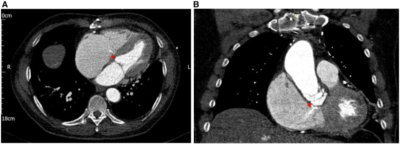
Figure 2.
(A) Transverse arterial phase-contrast computerized tomography image demonstrating a thin jet of contrast entering a dilated right atrium (red asterisk). (B) Coronal arterial phase-contrast computerized tomography image demonstrating a thin jet of contrast entering a dilated right atrium through a defect in the wall of the left ventricular outflow tract (red asterisk).

