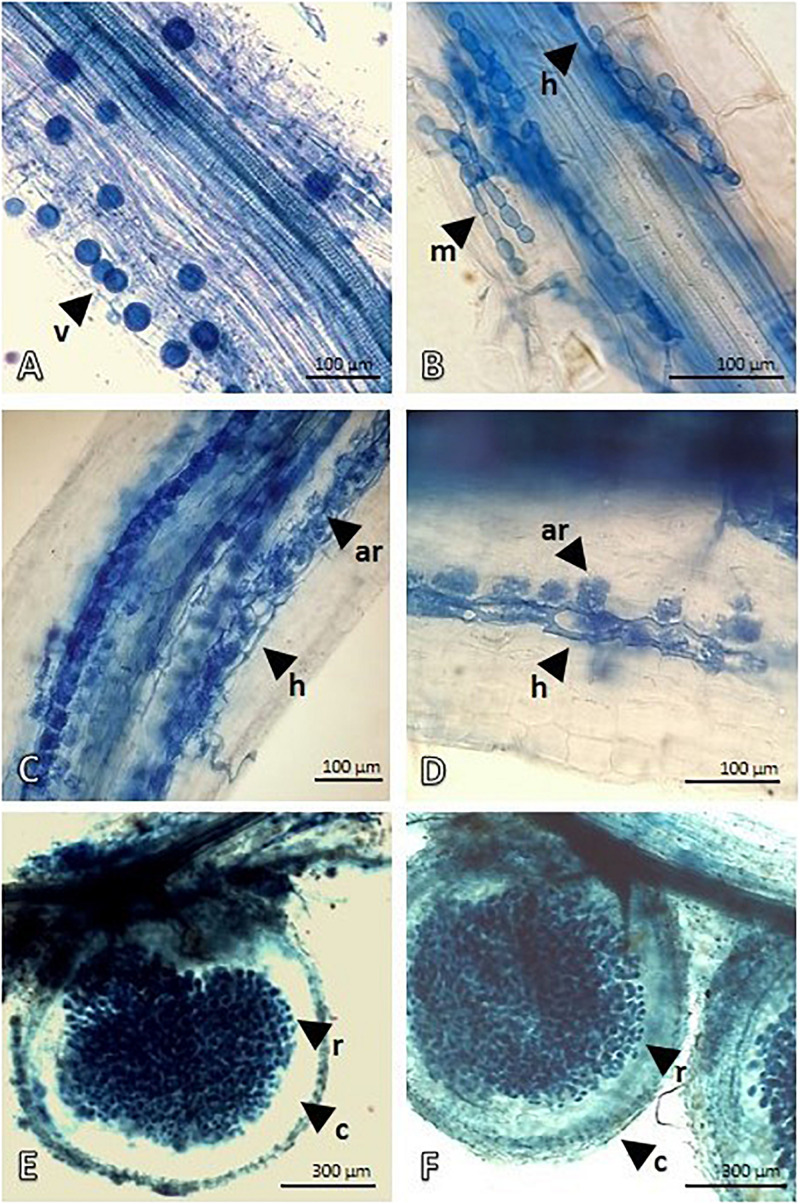
FIGURE 1.
Mycorrhization of Lotus and Festuca roots and Rhizobium nodules of Lotus roots detected by Trypan Blue staining and light microscopy. (A,B) show the mycorrhize colonization in Festuca roots. Intraradical vesicles (v) and hyphae (h) developing intracellular microsclerotia structure (m), with rounded hyphae cell, are visible in root fragments. (C,D) show Intraradical hyphae (h) depicting an advanced arbuscule colonization in Lotus roots. (E,F) display Lotus root nodules. Outer, lighter stained, cortex of nodule is well-distinguished from the symbiotic region that is heavily infiltrated by Rhizobium bacteria.

