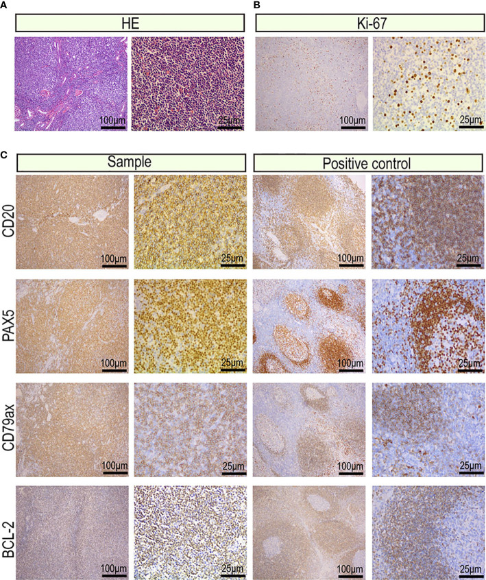
Figure 2.
Images of the tumor cells from the primary renal MALT lymphoma (scale bar: 100μm, 25μm). (A) Hematoxylin and eosin-stained tumor sections. (B) Representative IHC images for Ki-67 proliferation index of 20%–25%. (C) Representative IHC images for CD20, CD79a, PAX5 which were markers of B lymphocyte and BCL-2 which was a marker of non-germinal center lymphocyte.

