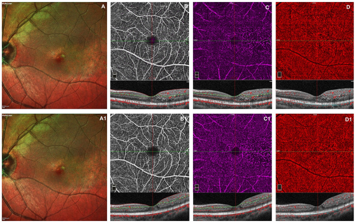
Figure 1.
Left eye of a male 20 years old patients before and after 3 monthly intravitreal injection of ranibizumab. At baseline, multicolor shows foveal exudates and retinal telangiectasia temporally to the fovea (A). OCTA images of the superficial capillary plexus (SCP) shows few areas of capillary hypoperfusion with a slight increased FAZ area (B). The deep capillary plexus (DCP) (C) shows irregularly dilatated small vessels, increased vascular rarefaction, in particular temporally the fovea. The CC reveals few areas of no flow signal due to a posterior shadow effect from intraretinal exudates (D). The structural spectral domain optical coherence tomography (SD-OCT) B-scan of each OCTAngiography (OCTA) image reveals some intraretinal exudates and cysts temporally the foveal region. After loading phase, the multicolor image reveals no change in foveal exudates and in teleangectasia temporally to the foveal region (A1). At OCTA no significant improvement in capillary drop out and anomalies in vessel size are found in superficial and deep capillary networks, respectively (B1,C1). In CC persists few areas of no flow signal due to the presence of intraretinal exudates (D1). No structural OCT changes was evident after treatment.

