Figure 1.
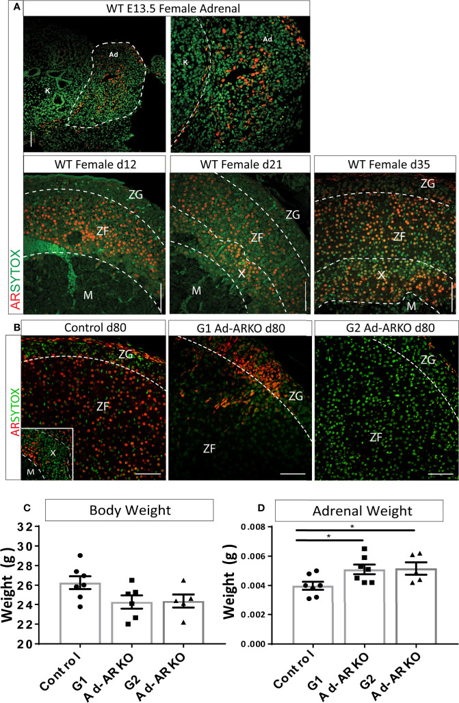
Confirmation of AR ablation in the adrenal cortex. (A) AR localization can be detected at e13.5 in the WT female adrenal, with staining localized to the central region of the fetal adrenal (N = 2). Immunostaining of AR during postnatal development shows localization from the inner most cortex/medulla boundary and ZF at d12 and d21. At d35, AR is observed in the X-zone, ZF and ZG (N = 3) (B) Immunostaining of AR in the adrenal cortex reveals partial ablation in the G1Ad-ARKO and complete adrenal AR ablation in the G2 Ad-ARKO females. Insert demonstrates AR localization in the X-zone of WT d80 Controls. Red: AR protein, Green: sytox counterstain. (C) Bodyweight analysis of d80 WT controls, G1Ad-ARKO and G2 Ad-ARKO females revealed no changes. (D) Adrenal weight analysis shows a significant increase in G1 and G2 Ad-ARKO mice compared to respective controls (one-way ANOVA; n=5-8, *p < 0.05, *p < 0.05, Tukey’s post-hoc analysis, error bars SEM). Annotations; X, X-zone; ZF, zona fasciculata; ZG, zona glomerulosa. Scale Bars 100 µm.

