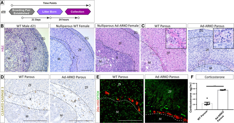
Figure 3.
AR is dispensable for X-zone regression during pregnancy. (A) Diagram depicting collections of G2 Ad-ARKO females and controls following pregnancy for X-zone regression analysis. Nulliparous G2 Ad-ARKO females and Nulliparous WT controls were placed in a breeding pair at d70. Females were collected 24 h post-partum. (B) X-zones are present in WT d21 males and WT virgin females. N=5. (C) Analysis of X-zone morphology following pregnancy in WT and G2 Ad-ARKO females reveals regression of the X-zone in both cohorts. N=5. (D) Immunostaining of cleaved caspase 3 shows apoptosis of the X-zone in both WT and G2 Ad-ARKO females. N=5. (E) Immunostaining of 20 α-HSD localization shows only a few X-zone cells remaining in the cortex of WT controls and G2 Ad-ARKO females. Red: 20alpha-HSD protein, Green: sytox counterstain. N=5. (F) Analysis of circulating corticosterone shows a significant increase in G2 Ad-ARKO females following pregnancy (Students t-test; n=4–6), ***p < 0.05, error bars SEM) compared to WT controls. Mice collected between d80-90. Scale Bars 100 µm. Annotations; M, medulla; X, X-zone; ZF, zona fasciculata; ZG, glomerulosa.

