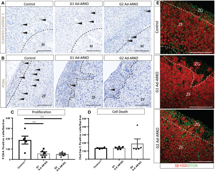
Figure 5.
Fewer proliferating cells are observed on G1 and G2 Ad-ARKO females compared to WT controls. (A, D) Investigation of apoptosis revealed no changes in either G1 or G2 Ad-ARKO females when compared to WT controls. Confirmation through cell counts reveals no changes in cell death in G1or G2 Ad-ARKO females compared to WT controls. (B, C) Immunostaining of PCNA localization and cell counts reveal fewer proliferating cells in G1and G2 Ad-ARKO females compared to WT controls. (E) Immunostaining of 3βHSD localization reveals that sections of the ZG do not express 3βHSD in G1 or G2 Ad-ARKO females, compared to WT controls that are positive in both the ZG and ZF. (*) denotes insert location. N=5. All mice collected and analyzed at d80. Scale Bars 100 µm. Annotations; M, medulla; ZF, zona fasciculata; ZG, zona glomerulosa.

